There are two types of donor tissues which are used in surgery. The sclera, or white portion of the eye; and the cornea, which is the clear lens at the front of the eye.
Corneal Transplants
The cornea is the clear lens at the front of the eye which focuses light onto the retina. Damage or disease to the cornea may cause it to change shape or become cloudy. This can cause pain, distorted or blurry vision. Corneal transplant replaces the damaged or diseased with a healthy donor cornea.
Penetrating Keratoplasty (PK)
The full thickness of the cornea is transplanted in this procedure.

Descemet’s Stripping Endothelial Keratoplasty (DSAEK), Descemet’s Membrane Endothelial Keratoplasty (DMEK)
These procedures are done to replace the Endothelial Layer of the cornea. The endothelial layer is a single cell layer at the back of the cornea. The endothelial cells act as pumps to maintain the pressure in the cornea and help keep it clear. If these cells do not function properly the pressure in the cornea will increase and it can become cloudy
Deep Anterior Lamellar Keratoplasty (DALK)
This procedure involved replacing the front layers of the cornea, the epithelium and stroma.

Sclera
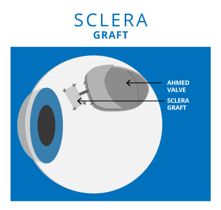
The sclera, or white portion of the eye, may be used in a number of different surgical procedures. Portions of the sclera, or grafts, are used. There can be up to eight sclera grafts from an eye donation.
Glaucoma Surgery
A graft, or patch, of sclera may be used over the tip of a pressure release valve (Ahmed Valve) during surgery for glaucoma.
Orbit Reconstruction
A prosthetic (artificial) eye may be implanted using donor sclera. The prosthetic is wrapped with sclera and the four muscles which move the eye are reattached to the donor sclera. This allows the eye to move in unison with the natural eye.
Eyelid Surgery
Donor sclera may be used in eye lid reconstruction.


